
Download
RESEARCH ARTICLE
Quality assessment of black ginseng materials utilizing chemometrics and modeling inflammation in Zebrafish
Lulu Wang1,2, Meijie Yu1,2, Yao Fu1,2, Li Li3,4, Xianghe Meng2,5, Jian Yang6,7, Min He1,2*, Mengmeng Sun1,2*
1Northeast Research Asia Institute of Traditional Chinese Medicine, Changchun University of Chinese Medicine, Jingyue Economic Development District, Changchun, China;
2The Jilin Province School-Enterprise Cooperation Technology Innovation Laboratory of Herbal Efficacy Evaluation Based on Zebrafish Model Organisms, Changchun University of Chinese Medicine, Jingyue Economic Development District, Changchun, China;
3Capital Medical University, Subsidiary Beijing Hospital of Traditional Chinese Medicine, Dongcheng District, Beijing, China;
4Beijing Institute of Traditional Chinese Medicine, Shuiche Alley Xinjiekou, Xicheng District, Beijing, China;
5Changchun Wish Technology Co., Ltd., Building E11, Area B, Beihu Science and Technology Park, High-tech North District, Changchun, China;
6State Key Laboratory of Component-based Chinese Medicine, Tianjin University of Traditional Chinese Medicine, Tianjin, China;
7Analytical Instrumentation Centre, Tianjin University, Tianjin, China
Abstract
Black ginseng is a novel variant of processed ginseng that is produced by subjecting ginseng to a series of nine steaming and drying cycles. However, the precise connection between the processing cycle and the quality of black ginseng remains uncertain, and further investigation is required to establish the association between quality markers and biological activity. In this research, high-performance liquid chromatography was employed to analyze the composition of 17 ginsenosides present in samples of black ginseng. The anti-inflammatory properties of black ginseng saponin extract were tested utilizing a zebrafish tail fin amputation model. The findings indicate that the concentrations of Rg3, Rg5, and Rk1 in black ginseng samples exhibited their highest levels following the seventh processing cycle, hence demonstrating the most effective regulatory impact on neutrophils and macrophages. This study posits that the correlation between these three ginsenoside and biological activities could potentially serve as a crucial factor in ensuring the quality control of black ginseng, thereby contributing to the advancement of quality control measures for health foods and dietary supplements that utilize black ginseng as their primary constituent.
Key words: black ginseng, ginsenoside, immune response, inflammatory models, quality assessment
*Corresponding Authors: Mengmeng Sun and Min He, Northeast Research Asia Institute of Traditional Chinese Medicine, Changchun University of Chinese Medicine, No. 1035, Boshuo Rd, Jingyue Economic Development District, Changc-hun 130117, China. Emails: [email protected]; [email protected]
Received: 25 September 2023; Accepted: 6 December 2023; Published: 22 January 2024
© 2024 Codon Publications
This is an Open Access article distributed under the terms of the Creative Commons Attribution-NonCommercial-ShareAlike 4.0 International (CC BY-NC-SA 4.0). License (http://creativecommons.org/licenses/by-nc-sa/4.0/)
Introduction
Ginseng, scientifically known as Panax ginseng C.A. Meyer, holds a prominent position as a well-recognized and extensively utilized herb globally (He et al., 2018). Its usage as an adaptogen, facilitating the recuperation, and prophylaxis of the human body, has been well--documented. Throughout the history of human utilization of ginseng, it has been observed that the hue of steamed ginseng materials, such as red ginseng, undergoes alterations (He et al., 2018). In addition, there exists sun-dried ginseng, which offers convenient preservation and acts as a preservative. Sugar ginseng is produced through maceration in honey. Fermented ginseng, on the other hand, undergoes a low-temperature fermentation procedure. Lastly, ginseng marinated in wine or vinegar is also a traditional way of processing. However, the process of steaming ginseng remains the most employed technique (In et al., 2017; Kim et al., 1985; Lim et al., 2010; Paek et al., 2006; Zhang and Xu, 2018). The steamed ginseng materials are deemed to be more conducive for preservation, and their physiological impacts have undergone alterations, particularly with the better modulation of the body’s immune system (He et al., 2018). Hence, the utilization of ginseng that undergoes steaming has consistently had significance as a drug in traditional medicine, while also serving as a crucial ingredient in numerous health foods and dietary supplements (Riaz et al., 2019).
Black ginseng is a novel variant of processed ginseng that is produced by subjecting ginseng to a series of nine steaming and drying cycles, resulting in a distinct black hue (Nam et al., 2012). The potential biological effectiveness of black ginseng, such as anti-tumor, anti-inflammatory, antioxidant, anti-aging, and anti-diabetic activities is believed to surpass that of ginseng products derived from other processing procedures (Huang et al., 2023). However, it is worth noting that black ginseng may exhibit increased cytotoxicity in laboratory settings, which could be attributable to the unique properties of certain saponin components present in black ginseng (Park et al., 2022). No apparent abnormalities were observed in the physiological indicators, blood biochemistry, and histopathology analyses following the administration of black ginseng extract, as determined through tests conducted on rat models (Park et al., 2022). The chemical composition of black ginseng, particularly ginsenosides, shows significant alterations during nine steaming and drying cycles, resulting in corresponding modifications to the biological activity of black ginseng (Huang et al., 2023). For instance, the repeated steaming and drying processing of black ginseng leads to an augmentation of its antioxidant properties (Kim et al., 2011). However, the technique of nine cycles of steaming and drying is derived from the conventional preparation procedures employed for certain herbs, such as Rehmannia glutinosa Libosch, in Traditional Chinese medicine (TCM) (Sun et al., 2018). At present, there exists a dearth of conclusive evidence about the correlation between the precise frequency of steaming and drying processes and the resultant quality and biological activity of black ginseng. In the production process, there is a deliberate reduction in the number of steaming and drying cycles for specific black ginseng materials. This reduction is motivated by factors such as cost, time efficiency, and material conservation. Thus, the presence of significant diversity in the biological activity of black ginseng materials available in the market is evident. Hence, the investigation of the correlation between the cycles of steaming and drying processes applied to black ginseng, and its resultant quality and efficacy, has emerged as a crucial concern in the realm of black ginseng product quality control.
In this research, the identification of the 17 ginsenosides present in the black ginseng material, which underwent a process of different cycles of steaming and drying, was conducted. Subsequently, a zebrafish inflammatory model was employed to evaluate the bioactivity of total ginsenoside extracts derived from different black ginseng materials. Here, transgenic zebrafish lines Tg (mpx:GFP/ mpeg:mCherry) were employed as a model system to investigate the impact of ginsenoside extracts on the migration of innate immune cells, specifically neutrophils and macrophages, during inflammatory conditions. Finally, we summarized a relationship between processing cycles and the quality and anti-inflammatory efficacy of black ginseng. Our findings contribute to the understanding of quality control measures for black ginseng and offer novel research evidence for enhancing the overall quality of black ginseng products.
Materials and Methods
Preparation of black ginseng materials
Raw materials: The raw roots and rhizome of Panax ginseng C.A. Meyer (5-year-old) were obtained from the Beijing Institute of Traditional Chinese Medicine in Beijing, China. Before submitting to Changchun University of Chinese Medicine in Changchun, China, Prof. Dr. Li Li validated all samples. To prepare processed black ginseng, the raw materials were subjected to drying in an oven at a temperature of 50°C. Subsequently, the dried materials were steamed with water for 180 min at 100°C (Metwaly et al., 2019). Following this, the processed materials were dried at the same temperature of 50°C for 24 h (Metwaly et al., 2019). The production of the carcinogen benzo(a)pyrene is an inevitable consequence of the distinctive heat treatment procedure employed in the preparation of black ginseng. The concentration of this hazardous compound will increase rapidly with an increase in the drying temperature. Hence, to mitigate the excessive carbonization of black ginseng, a drying temperature of 50°C is employed (Jo et al., 2009). This process was repeated nine times to obtain black ginseng materials corresponding to each cycle from one to nine, and the dried product of fresh ginseng was used as the control group.
Preparation of ginsenoside extracts from black ginseng materials
The methodology employed for the extraction of ginsenoside from black ginseng samples, which have undergone zero to nine cycles of preparation, adheres to the guidelines outlined in the Pharmacopoeia of the People’s Republic of China (2020) (Commission, 2020). In accordance with the specifications for experimental design, the extraction process was optimized as follows: the black ginseng samples’ roots and rhizome were pulverized, and a precise measurement of approximately 2 g of black ginseng powder was obtained. This powder was then placed into a 50 mL volumetric flask, followed by the addition of 50 mL of chloroform. The mixture was allowed to stand overnight and subsequently subjected to ultrasonic degreasing for 1 h. The resulting chloroform liquid was discarded, and the solvent of the residual drug was evaporated. The remaining substance was transferred into a 100 mL Erlenmeyer flask along with filter paper. The experimental procedure involves the addition of precisely 50 mL of water-saturated n-butanol into a container. The container should be securely sealed and stored. Following an overnight period, the mixture should be subjected to sonication for 30 min, utilizing a power of 250 W and a frequency of 50 kHz. The primary filtrate should be filtered and discarded. Then, a precise volume of 25 mL of the continuous filtrate was transferred to a test tube. The test tube was subjected to a nitrogen blow drier to facilitate drying. The resulting residue in the test tube was dissolved immediately in methanol to achieve a test solution concentration of 40 mg/mL. The solution should be thoroughly shaken. The extraction solution underwent filtration using a 0.22 μm filter membrane and was thereafter introduced into the high-performance liquid chromatography (HPLC) system through direct injection.
Ginsenoside reference standards preparation
The 17 reference samples were used as standards at different concentrations to quantify the amounts present in the processed samples. Monomeric ginsenoside Rb1 (3.008 mg/mL), Rb2 (1.508 mg/mL), Rb3 (2.561 mg/mL), Rc (2.012 mg/mL), Rd (1.308 mg/mL), Re (1.520 mg/mL), Rf (1.610 mg/mL), Rg1 (2.412 mg/mL), Rg2 (2.068 mg/mL), Rg3 (0.442 mg/mL), Rg5 (0.173 mg/mL), Rh1 (0.772 mg/mL), S-Rh2 (0.281 mg/mL), R-Rh2 (0.428 mg/mL), Rk1 (0.242 mg/mL), Ro (2.521 mg/mL), and F1 (1.825 mg/mL) were dissolved in methanol and diluted to obtain a series of mixed standard solutions of different concentrations. The above standards were acquired from Beijing Solarbio Science & Technology Co., Ltd, located in Beijing, China. The purity of all reference substances exceeded 98%.
High-performance liquid chromatography analysis
The HPLC analysis was conducted utilizing an Agilent 1260 instrument (Agilent Technologies, Singapore International Pte. Ltd). The equipment was ensembled with a micro-vacuum degasser, quaternary pump, auto-sampler, thermostated column compartment, and diode array detector. The diode array detector was connected to an Agilent ChemStation. The experiment involved the utilization of an Agilent 5 µm-ZORBAX SB-C18 column (4.6 × 250 mm; USA) for conducting chromatographic analysis. The column was kept at a constant temperature of 40°C (Sun et al., 2009). The wavelength of detection utilized in the experiment was 203 nm. The binary gradient elution system utilized a mixture of acetonitrile, water, and a 5% acetic acid aqueous solution (in a ratio of 10:85:5, v/v/v) as solvent A, and a mixture of acetonitrile and water (in a ratio of 80:20, v/v) as solvent B (Sun et al., 2009). The separation was accomplished by implementing a gradient program consisting of the following conditions: at 0 min, the mobile phase composition of 0% B was employed; from 0 to 10 min, the mobile phase composition was adjusted to 30% B; from 10 to 25 min, the mobile phase composition was further modified to 50% B; from 25 to 40 min, a mobile phase composition of 100% B was utilized; from 40 to 50 min, the mobile phase composition remained at 100% B; from 50 to 53 min, the mobile phase composition was reverted to 0% B; and finally, from 53 to 60 min, a mobile phase composition of 0% B was maintained (Sun et al., 2009). The flow rate was established at a rate of 1.5 mL per minute, whereas the sample injection volume was 10 mL. A methanol-based solution was created, consisting of 17 bioactive reference chemicals. The employed methodology for analyzing the chemical contents is widely recognized and has been thoroughly validated, demonstrating satisfactory levels of reproducibility and repeatability for each ingredient. The limit of detection (LOD), the limit of quantification (LOQ), and R2 values are listed in Table S1.
Zebrafish tail-fin amputation model for anti-inflammatory tests
The induction of local inflammation in zebrafish can be achieved by performing tail-fin amputation on zebrafish larvae (He et al., 2020). The present model can be utilized for the examination of the immunomodulatory response by investigating the migratory behavior of macrophages and neutrophils (Xie et al., 2021). In this study, zebrafish larvae of double transgenic lines Tg (mpx:GFP/mpeg:mCherry) were used as the research object, and tail-fin amputation was exploited to explore the migration patterns of neutrophils and macrophages. To track the movement of neutrophils, the myeloid-specific peroxidase (mpx) was genetically modified to express a green fluorescent protein (GFP). Similarly, the migration of macrophages was indicated by marking the -macrophage-expressed gene (mpeg) with a red fluorescent mCherry protein.
Study on identifying the suitable ginsenoside concentration: The culture medium for zebrafish, known as egg water, is prepared by combining distilled water with a certain proportion of quick sea salt. Ginsenoside extracts were dissolved in egg water to prepare solutions with concentrations of 20 μg/mL, 50 μg/mL, and 100 μg/mL, respectively. Zebrafish larvae at the age of three days were immersed in a solution of egg water that included varying concentrations of ginsenoside extract for 96 h. During this time frame, the researchers conducted an observation of the fatal and teratogenic effects of ginsenoside extract on zebrafish at intervals of 24 h, 48 h, 72 h, and 96 h. The purpose of this inquiry is to provide further elucidation on the safe concentration of ginsenoside extract.
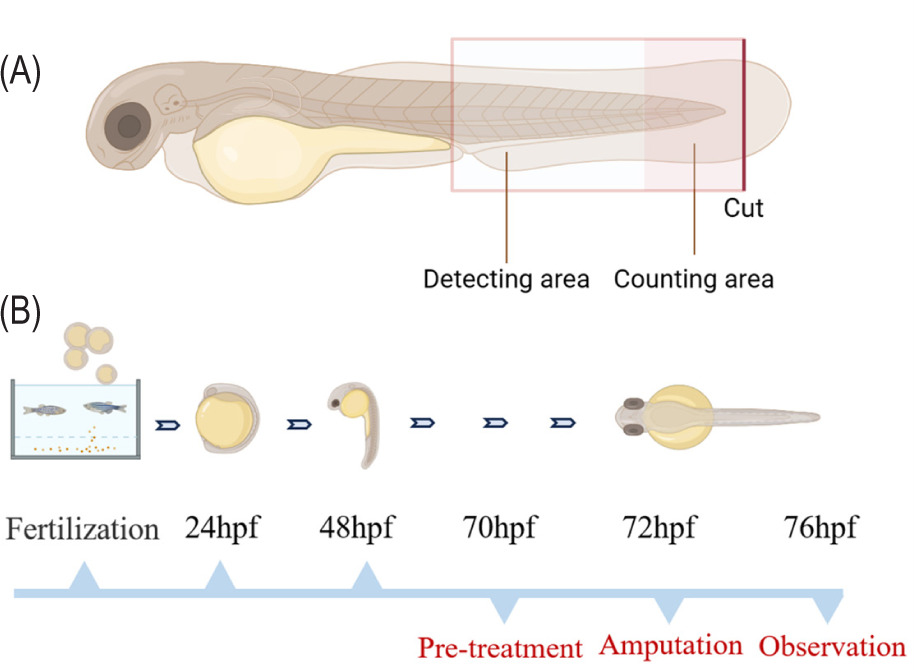
Immunomodulatory study of ginsenoside on zebrafish: Zebrafish larvae that were three days old were subjected to a pretreatment period of 2 h. During this period, they were exposed to either a solution containing ginsenoside extract at a concentration of 20 μg/mL or beclomethasone or egg water. This pretreatment was conducted before the amputation procedure (He et al., 2020; Xiong et al., 2022). Next, the larvae were relocated into a solution of egg water supplemented with 0.02% tricaine (Yuanye Bio-Technology Co., Ltd, Shanghai, China) to induce anesthesia. Following this, the larvae were arranged in Petri dishes that were coated with a layer of 2% agarose. This setup facilitated the subsequent process of partially amputating the tail-fin, as depicted in Figure 1A. The amputation procedure was performed utilizing a Leica M165C stereomicroscope, a micromanipulator, and a 1 mm sapphire blade manufactured by World Precision Instruments (He et al., 2020; Xiong et al., 2022). Following that, the zebrafish larvae that had undergone amputation were then reintroduced into either a fresh solution of ginsenoside extract or egg water for 4 h, as depicted in Figure 1B. Zebrafish larvae were subjected to microscopic examination for real-time observation to facilitate the examination of macrophage and neutrophil migration.
Figure 1. The provided diagram illustrates the experimental setup for tail-fin amputation tests conducted on -zebrafish. (A) The term “detecting area” refers to the visible area observed under a microscope, while the term “counting area” refers to the specific region utilized for counting migrating cells. The thick red line is used to show the place of amputation. (B) The time series of the experiment.
The zebrafish used in this study were cared for and managed in accordance with the standards provided by the Zebrafish Model Organism Database (http://zfin.org) and in adherence to the regulations set forth by the local animal welfare council of Changchun University of Chinese Medicine. All animal procedures were conducted in strict adherence to the Guidelines for Care and Use of Laboratory Animals established by Changchun University of Chinese Medicine. The experiments were duly authorized by the Animal Ethics Committee of Changchun University of Chinese Medicine (Approval No. 2020233).
Data processing and statistical analysis
The analysis involved the determination of the concentration of 17 components present in each extract of ginsenoside. The K-means clustering approach was employed to differentiate between the 17 chemicals present in ginsenoside extracts. This analysis was conducted using the tools included in the MetaboAnalyst 5.0 software program (Pang et al., 2022), which can be accessed at http://www.metaboanalyst.ca. Subsequently, a two-tailed, unpaired Student’s t-test was conducted using SPSS version 23.0 (IBM, Armonk, NY) to compare the various ginsenoside extracts based on the quantities of each detected compound and the overall amounts of these 17 compounds, respectively. Statistical significance was determined at a significance level of p < 0.05.
The imaging of severed larvae was conducted utilizing a Leica MZ16FA fluorescence stereomicroscope, with support of LAS version 3.7 software. The quantification of migrating neutrophils with green color and macrophages with red color was performed on the wound site of a zebrafish larva, as depicted in Figure S1B. Subsequently, a one-way analysis of variance (ANOVA) was employed, followed by a post hoc analysis using the least significant difference (LSD) method, to compare the cell counts between the groups subjected to amputation and those treated with ginsenoside extract. Statistical significance was determined at a significance level of p < 0.05.
Results
Chemical analysis of ginsenoside extracts
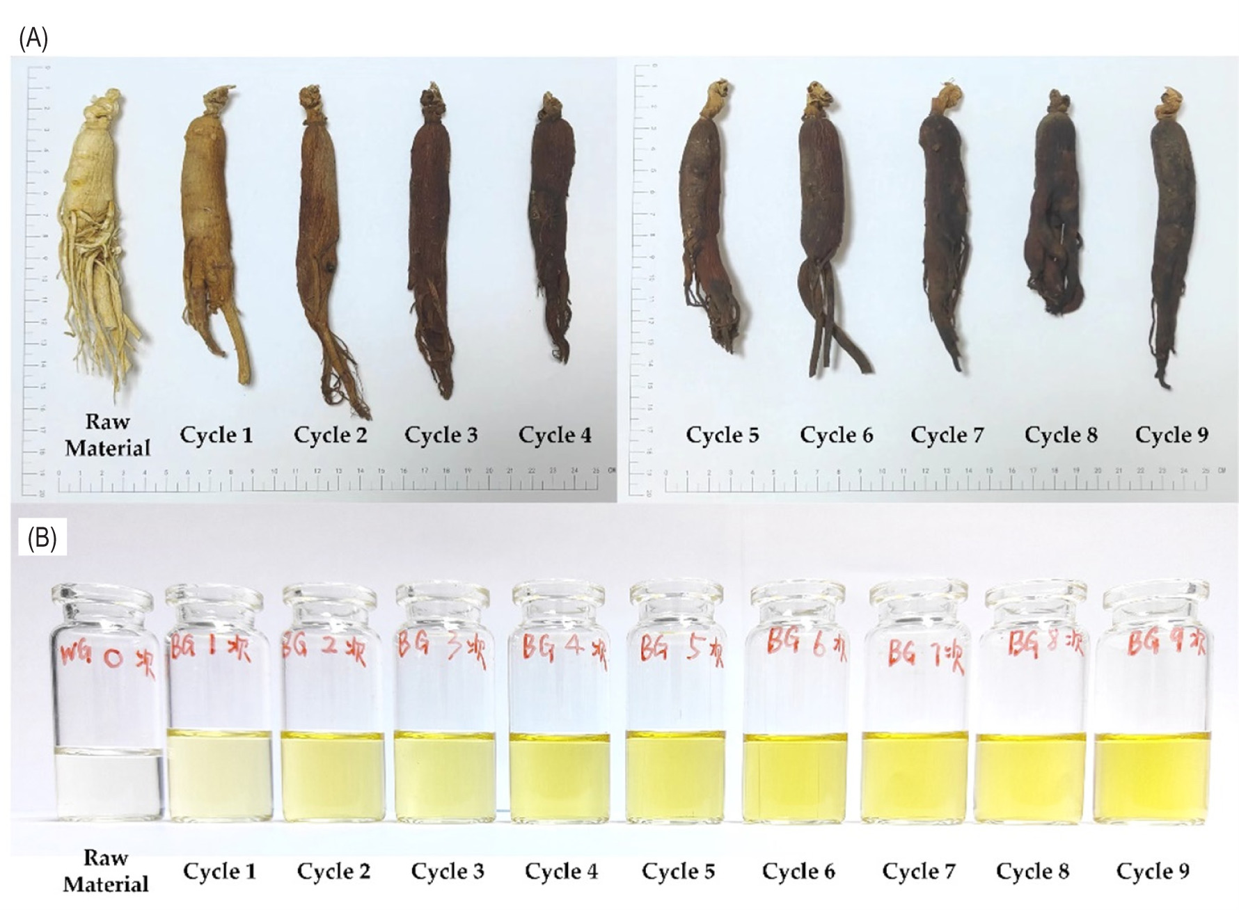
The process of preparing black ginseng involves a repeated cycle of steaming and drying, which is performed nine times. Throughout each steaming and drying cycle, samples of the black ginseng are carefully preserved. The observation depicted in Figure 2A illustrates that with an increase in the processing cycle, there is a steady darkening of the color of the black ginseng sample. Similarly, Figure 2B demonstrates a corresponding darkening of the color of the ginsenoside extract.
Figure 2. Samples of black ginseng and ginsenoside extract obtained from various processing cycles. (A) Raw and black -ginseng samples. (B) Ginsenoside extract samples.
To evaluate the chemical characteristics of ginsenoside extracts, the concentrations of the 17 main ginsenoside components were quantified in the extracts derived from black ginseng samples. These samples were taken at various cycles during the steaming and drying process, and three distinct batches of black ginseng samples were acquired during each processing cycle. Hence, a comprehensive set of 30 samples was identified for the particular analysis of 17 ginsenosides utilizing HPLC.
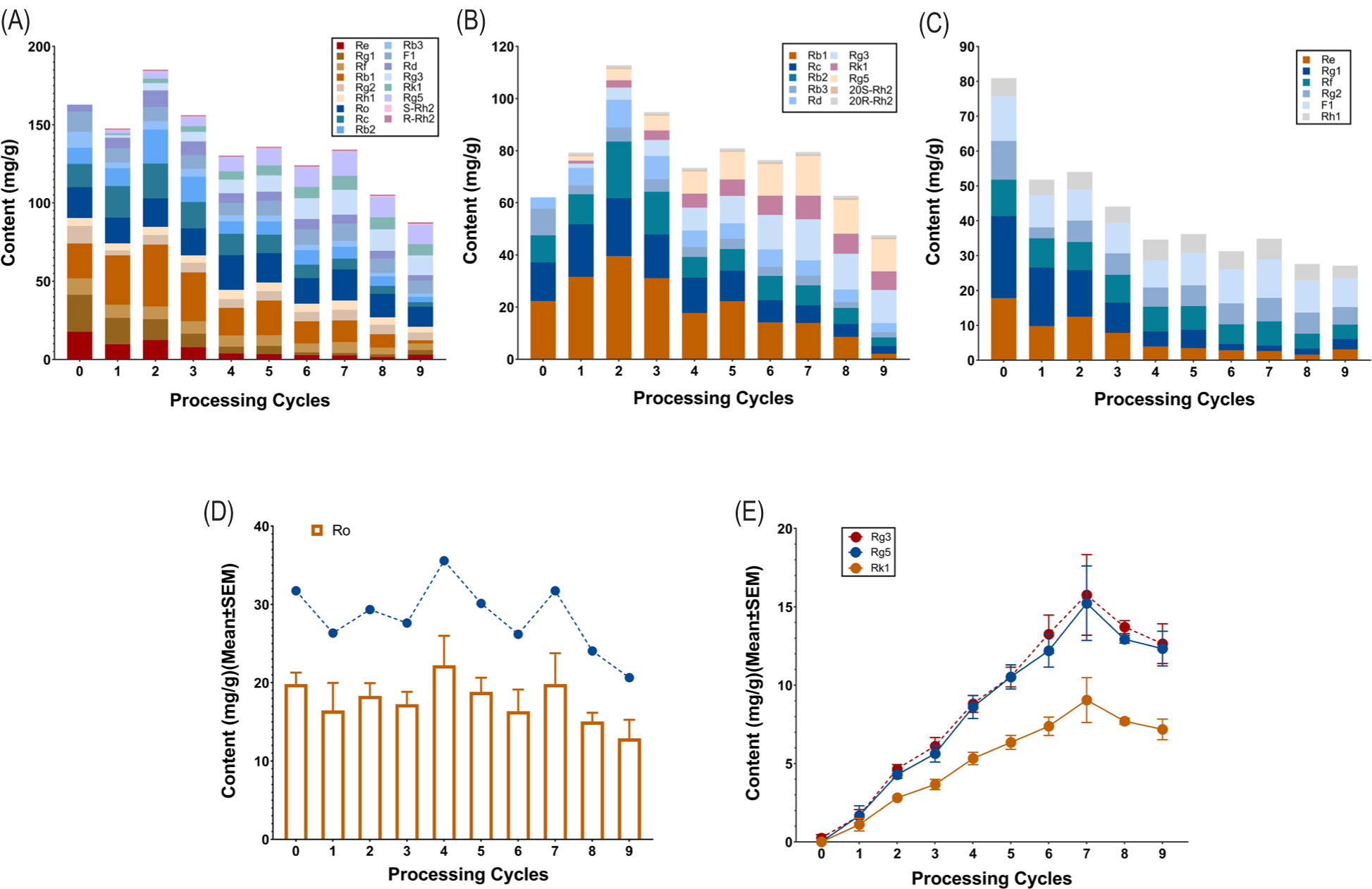
Upon consolidating the chemical data from three batches of black ginseng samples across each processing cycle, it is evident from Figure 3A that the cumulative concentration of the 17 detected ginsenoside in the black -ginseng samples generally exhibited a declining pattern as the processing cycle increased. Ginsenosides, which are -triterpenoid glycosides, can be categorized into various types according to the structure of the aglycone. These types include protopanaxadiols (e.g., monomeric ginsenoside Rb1, Rb2, Rb3, Rc, Rd, Rg3, Rg5, Rk1, and Rh2), protopanaxatriols (e.g., monomeric ginsenoside Re, Rg1, Rg2, Rf, F1, and Rh1), and oleanane (e.g., monomeric ginsenoside Ro) (Elshafay et al., 2017; Wei et al., 2020). The results depicted in Figure 3B, 3C, and 3D indicates a general decline in the levels of the three distinct configurations of ginsenosides as the processing cycle increases. However, it is worth noting that this decreasing trend does not hold for ginsenosides Rg3, Rg5, and Rk1. The levels of these three individual ginsenosides exhibited a notable increase as the processing cycle progressed, ultimately reaching their peak after the seventh processing cycle, followed by a subsequent decline (Figure 3E).
Figure 3. The dynamic variations in the composition of 17 ginsenosides over nine processing cycles. (A) The chemical profiles of the detected 17 ginsenosides. (B) The chemical profiles of the detected ginsenoside of protopanaxadiols type. (C) The chemical profiles of the detected ginsenoside of protopanaxatriols type. (D) The chemical profiles of the detected ginsenoside of oleanane type (e.g., monomeric ginsenoside Ro). (E) The dynamic variations in the composition of Rg3, Rg5, and Rk1. Note: Processing cycle 0 indicates raw dried ginseng materials.
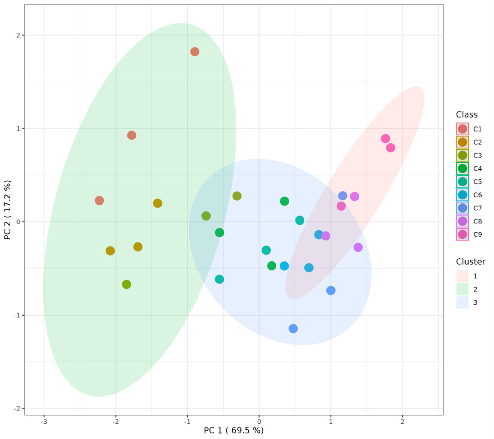
Subsequently, an unsupervised K-means clustering methodology (Likas et al., 2003) was employed to analyze the chemical data, to visually represent the variations among distinct ginsenoside extracts derived from samples of black ginseng. The results of K-means clustering are depicted in Figure 4. When the number of groups was set to three, all 27 samples of black ginseng were continuously allocated to distinct clusters. The details of K-means clustering are listed in Table 1. The black ginseng samples acquired following the third and seventh processing cycles serve as the demarcation points that delineate the three clusters. Hence, we choose to collect samples from the aforementioned two demarcation points, as well as distinct black ginseng samples within each cluster (namely, samples derived from cycles 1, 3, 4, 7, and 9) for further analysis.
Figure 4. K-means clustering results based on the content of 17 ginsenosides in black ginseng samples collected from nine distinct processing cycles. C means processing cycle.
Table 1. The detailed results of K-means clustering analysis.
| Cluster | Members |
|---|---|
| Cluster 1 | C7-S2; C8-S1; C8-S2; C8-S3; C9-S1; C9-S2; C9-S3 |
| Cluster 2 | C1-S1; C1-S2; C1-S3; C2-S1; C2-S2; C2-S3; C3-S1; C3-S2 |
| Cluster 3 | C3-S3; C4-S1; C4-S2; C4-S3; C5-S1; C5-S2; C5-S3; C6-S1; C6-S2; C6-S3; C7-S1; C7-S3 |
Note: C – processing cycle; S – specific black ginseng sample.
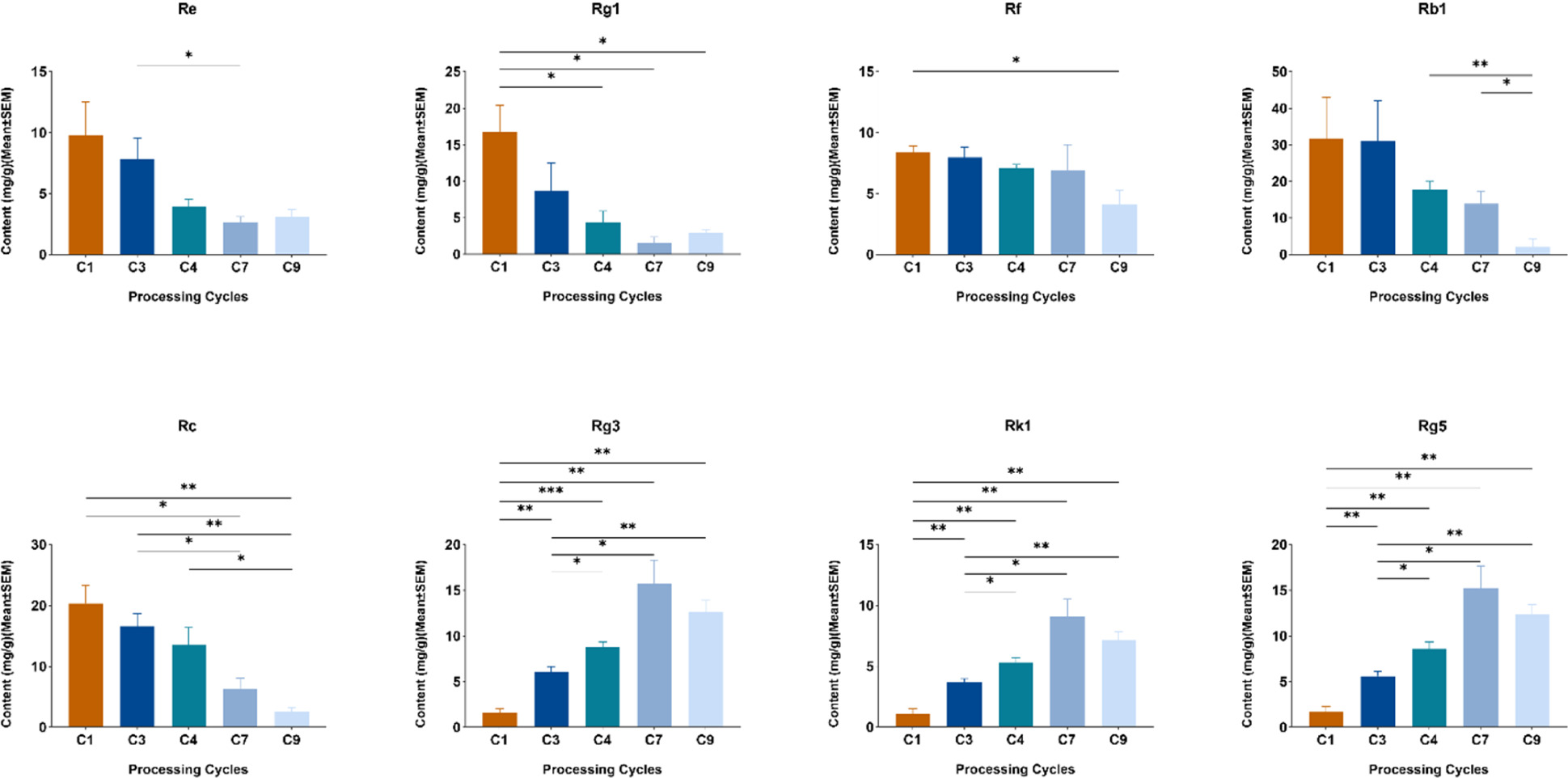
Next, ANOVA was employed to assess the levels of 17 ginsenosides among samples of black ginseng that underwent different processing cycles, specifically cycles 1, 3, 4, 7, and 9. The findings indicated that there were notable variations in the levels of monomeric ginsenosides Rg1, Rb1, Rc, Re, Rf, Rg3, Rg5, and Rk1 among the various black ginseng samples (Figure 5). Noteworthy are the ginsenosides Rg3, Rg5, and Rk1. The contents of these three chemicals in the samples following the first and third processing cycles exhibited notably lower values compared to the outcomes seen in the remaining processing cycles (p < 0.05) (Figure 5). Furthermore, whereas the black ginseng sample following the seventh processing cycle exhibits the highest concentration of these three components, there is no discernible difference when compared to the samples acquired during the fourth and ninth processing cycles (Figure 5).
Figure 5. The histograms compare the levels of certain ginsenoside compounds across different cycles, including cycles 1, 3, 4, 7, and 9. *p < 0.05; **p < 0.01; ***p < 0.001.
Anti-inflammatory activity tests
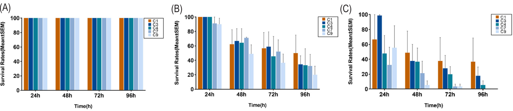
In this study, three different concentrations of ginsenoside extract obtained from black ginseng samples were administered to zebrafish larvae at the age of 3 days. The ginsenoside extract was derived from three separate samples collected during processing cycles 1, 3, 4, 7, and 9, respectively. The administration of the extract was done by introducing it into the egg water of the cultured zebrafish larvae. The exposure duration lasted for 96 h. During the specified time frame, the replacement of new egg water, which contained ginsenoside extract at a consistent dosage, occurred every 24 h. The survival rate of 30 zebrafish larvae was subsequently recorded for each experimental group. The experimental findings indicate that zebrafish larvae exhibited 100% survival when exposed to ginsenoside extracts at a dosage of 20 μg/mL (Figure 6A). However, it was observed that as the concentration of ginsenoside extract was augmented, there was a notable rise in the death rate of zebrafish larvae, particularly at a concentration of 100 μg/mL (Figure 6C). Hence, a concentration of 20 μg/mL of ginsenoside extract was chosen to evaluate the anti-inflammatory efficacy of black ginseng.
Figure 6. Safety evaluation results of ginsenoside extract. (A) The concentration of ginsenoside extract is 20 μg/mL. (B) The concentration of ginsenoside extract is 50 μg/mL. (C) The concentration of ginsenoside extract is 100 μg/mL.
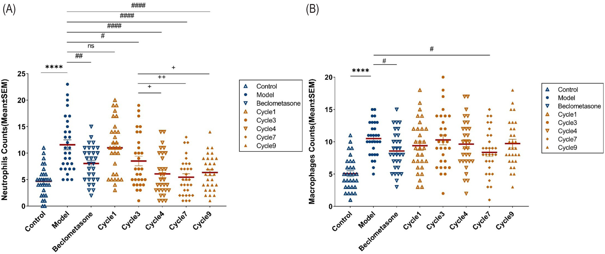
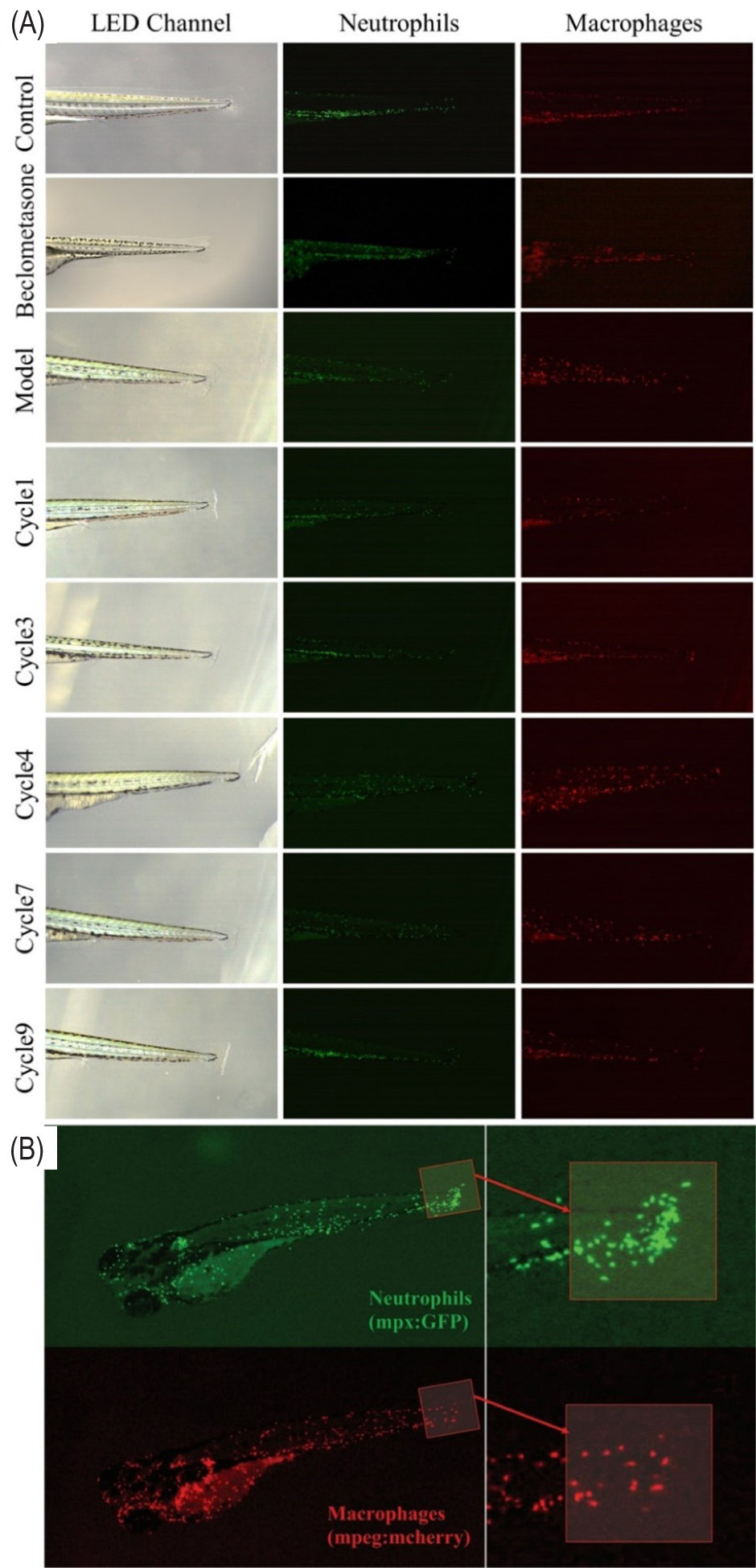
In order to assess the anti-inflammatory properties of ginsenoside extracts, a conventional zebrafish tail-fin amputation paradigm was employed. The ginsenoside extracts from three independent black ginseng samples obtained during each processing cycle were mixed, and afterward, the ginsenoside extracts (20 μg/mL) were examined for their anti-inflammatory activity based on the regulating effect of neutrophils and macrophages during the formation of inflammatory lesions in the tail of zebrafish larvae (Figure S1 A). The findings indicate that following injury to the tail-fin of zebrafish larvae, there was a substantial increase in the population of neutrophils and macrophages in the tail, indicating a pronounced inflammatory response (Figure 7). Furthermore, the administration of the positive drug (beclomethasone) demonstrated efficacy in reducing the abundance of neutrophils and macrophages in the tail (Figure 7).
Figure 7. The enumeration of migrating macrophages and neutrophils. (A) Neutrophils. (B) Macrophages. *Control versus Model, *p< 0.05, ****p< 0.0001; # Model versus Treatment, # p< 0.05, ## p< 0.01, #### p< 0.0001; + Comparison of different cycles, +p< 0.05, ++p< 0.01; ns, no significant difference.
The ginsenoside extract derived from black ginseng samples that were processed in the first cycle did not exhibit a statistically significant impact on neutrophil count (Figure 7A). Conversely, the ginsenoside extract derived from black ginseng samples that were processed in the third cycle demonstrated a notable down-regulating effect on neutrophil count (p < 0.05) (Figure 7A). Additionally, the ginsenoside extract derived from black ginseng samples that were processed in the fourth, seventh, and ninth cycles displayed a significant down-regulating effect on neutrophil count (p < 0.05), surpassing the down--regulating effect observed in the samples derived from the third processed cycle (p < 0.05), even superior efficacy compared to the positive drug (Figure 7A). Moreover, it is noteworthy that alone the ginsenoside extract subjected to the seventh processing cycle exhibited a substantial down--regulatory impact on macrophage count (p < 0.05) (Figure 7B). This effect closely resembled that of the positive drug. The other ginsenoside extracts did not have a discernible modulatory impact on the accumulation of macrophages at inflammatory locations (Figure 7B). In summary, the saponins derived from black ginseng, when obtained during the seventh processing cycle, exhibit a heightened capacity to modulate immune cells more completely.
Discussion
The initial preparation of black ginseng took place in South Korea, after which its usage spread extensively to China and many Southeast Asian nations (Metwaly et al., 2019). Throughout this study, individuals employed a multitude of processing techniques (Metwaly et al., 2019). The utilization of the nine-time steaming and drying method in this study is based on its historical significance as a traditional technique for processing herbs in TCM (Wang et al., 2009). However, regardless of the type of processing technique employed, the temperature and duration of steaming and drying are the most crucial factors to consider (Jo et al., 2014). The ginsenoside profile of ginseng underwent significant alterations as a result of processing aimed at achieving a black coloration, both externally and internally, as evidenced by chemical analysis (Nam et al., 2012). The level of browning in ginseng increases as the intensity of steaming treatment rises, primarily due to the reduction in moisture content and the subsequent carbonization resulting from repeated cycles of steaming and drying. Simultaneously, the hydrolysis and decomposition of sucrose occurs, resulting in the formation of glucose and fructose. During successive steaming cycles, the interaction between glucose, fructose, and free amino acids leads to a chemical reaction known as the “Maillard reaction” (Jin et al., 2015; Kim et al., 2013; Oh et al., 2021). This reaction occurs when the carbonyl group of the reducing sugar reacts with the free amino group of the amino acid, resulting in browning. Nevertheless, the elevation in temperature and the frequency of processing instances lead to the occurrence of a carbonization reaction in ginseng, resulting in the generation of the hazardous compound known as benzo(a)pyrene (Jo et al., 2009). Hence, the examination of black ginseng holds significance not only in relation to its biological efficacy but also necessitates comprehensive investigation into its safety aspects.
However, quantitative testing on benzopyrene was not conducted in this study for the following reasons: One aspect of our study primarily centered on investigating the impact of steaming and drying cycles on the immunomodulatory properties of black ginseng. On the other hand, drawing from prior research, the steaming and drying temperatures utilized in our experiment resulted in a lower level of benzopyrene, falling well within the established safety thresholds (Jo et al., 2009; Lee and Shim, 2007). Moreover, when considering the practical utilization of ginseng in the context of healthy food, it is advised to consume 2 to 3 g of dried plant material or 3 to 9 g of tea dosage daily (Yennurajalingam et al., 2015). However, it is worth noting that there is a dearth of comparable evaluations regarding the safe dosage of black ginseng, and the potential impacts of benzopyrene at this dosage remain uncertain. This implies that the comprehensive assessment of the functional properties of active constituents and the safety concerns associated with toxic constituents following the processing of black ginseng remains challenging at present. Hence, further research is warranted to do a comprehensive and thorough examination of safety in the future.
Ginsenosides are the primary biologically active compounds found in ginseng (Ratan et al., 2021). Several pharmacopeias in various nations and regions have designated certain markers, such as Rg1, Rb1, and Re, as indicators of high content and quality in ginseng (Commission, 2016; Commission, 2020; Committee, 2011; Committee, 2013; Convention, 2000). However, our analysis reveals that the levels of those monomeric ginsenosides in black ginseng exhibit a notable drop as the processing cycle increases. In contrast, the concentrations of Rg3, Rg5, and Rk1 experience a considerable increase, which is consistent with prior research findings (Jo et al., 2014). Following the application of heat treatment to ginseng, an internal denaturation reaction is expected to take place (Chen et al., 2020). The malonyl residues present in the molecular structure of protopanaxadiol saponins Rb1, Rc, Rb2, and Rd exhibit thermal instability and are susceptible to hydrolysis upon exposure to steam. This hydrolysis process leads to the conversion of these malonyl residues into Rg3. Following the process of dehydration, a conversion occurs resulting in the formation of Rg5 and Rk1 from Rg3 (Lee et al., 2015). The initial concentration of typical saponins found in ginseng tends to diminish after successive processing, while the proportion of infrequent saponins formed because of events such as demalonylation, deglycosylation, and hydrolysis tends to rise (Piao et al., 2020). This implies that future investigations should thoroughly examine the potential impacts of Rg3, Rg5, Rk1, and other relevant factors on the quality assurance of black ginseng.
Recently, scientists have put forth the notion that the therapeutic regulation of herbs and their extracts ought to encompass more than just the identification of key compounds. They argue that it is equally important to assess their biological activities and develop a quality evaluation framework capable of elucidating the correlation between indicators and activities (Kang et al., 2019). The primary biological activity of ginseng lies in its capacity to modulate the immune system (He et al., 2018). Consequently, an anti-inflammatory assessment was conducted using the zebrafish tail-fin amputation model to investigate the impact of black ginseng saponins on the functionality of innate immune cells. This is to get a deeper understanding of the variances in biological activity among black ginseng samples derived from various processing cycles. Furthermore, the utilization of transgenic zebrafish has proven to be a potent methodology for investigating the anti-inflammatory and immunomodulatory properties of ginsenoside extracts and monomeric ginsenosides, allowing rapid and visual evaluation (Sun et al., 2020). Based on the chemical analysis and bioactivity analysis conducted in this study, it can be inferred that there is a positive correlation between the duration of the processing cycle and the levels of Rg3, Rg5, and Rk1. Once the concentrations of the three components have reached a specific threshold, namely at the completion of the fourth cycle, the discernible disparity in the regulatory efficacy of black ginseng saponin extracts on neutrophils becomes less apparent. However, it is only when the concentrations of Rg3, Rg5, and Rk1 reach their maximum levels, specifically after the completion of the seventh cycle, that the saponin extract derived from black ginseng will exert a notable influence on the activity of macrophages. This observation suggests that there may be a positive relationship between the combined levels of Rg3, Rg5, and Rk1 present in the extract and its capacity to modulate the immune system. While it is acknowledged that other ginsenoside components may also influence the immunomodulatory activity of the samples (He et al., 2020), our research demonstrates the correlation between the processing cycle and the content and biological activity of the three aforementioned ginsenosides. Furthermore, many reports have revealed that Rg3, Rg5, and Rk1 exert an influence on neutrophils or macrophages during the anti-inflammatory process (Heo et al., 2023; Kang et al., 2018; Kim et al., 2012; Lee et al., 2013b; Shen et al., 2017; Xiong et al., 2022; Yu et al., 2017).
Rg3, Rg5, and Rk1 exhibit diverse pharmacological properties, encompassing anti-tumor effects (Choi et al., 2015), immune regulatory capabilities (Yoo et al., 2021), and the potential to ameliorate kidney injury through the modulation of inflammation and apoptosis (Park et al., 2015). Prior research has indicated that the potential mechanism underlying the whitening effects of ginsenosides Rg5 and Rk1 could include the activation of the MEK-ERK signaling pathway (Jin et al., 2018). Furthermore, it has been observed that the inhibition of TGF-β1-induced epithelial-mesenchymal transition and the promotion of stem-like characteristics are achieved through the downregulation of Smad2/3, NF-kB, ERK, p38 MAPK, and JNK signaling pathways in non-small cell lung cancer by Rk1 and Rg5 (Kim et al., 2021). The administration of Rg3 resulted in a reduction of the upregulation of pro--inflammatory markers, including tumor necrosis factor (TNF)-α, interleukin (IL)-1β, and cyclooxygenase (COX-2), within the hippocampus (Lee et al., 2013a). Apoptosis was observed to be produced by Rg3, which was found to activate caspase-3, caspase-8, and caspase-9, while also modulating the expression of Bcl-2 and Bax (Park et al., 2014). In addition, the administration of Rg5 resulted in the inhibition of TNF-α and IL-1β production, as well as a decrease in the expression of NF-κB, p65, and COX-2 in mice with renal inflammation induced by cisplatin (Li et al., 2016). Similarly, the administration of Rk1 demonstrated a considerable inhibitory effect on the excessive production of TNF-α and IL-1β, as well as a reduction in the expression of inducible nitric oxide synthase and COX-2 in hepatic tissues of mice (Hu et al., 2019). Furthermore, it should be noted that Rk1 exhibits inhibitory effects on NMDA receptors, thus presenting itself as a potentially valuable therapeutic intervention for pathological conditions associated with neuronal excitotoxicity (Ryoo et al., 2020). Pharmacokinetic studies reveal a notable finding, indicating that Rg3, Rg5, and Rk1 exhibit superior rates of absorption compared to Rb1, Rb2, and Rc (Yoo et al., 2021). This presents compelling evidence that Rg3, Rg5, and Rk1 exert their effects on pathways associated with inflammation and the immune response, underscoring the significant biological impact of these ginsenoside molecules. Hence, it is postulated that the Rg3, Rg5, and Rk1 could serve as significant indicators in the realm of black ginseng quality control, particularly in enhancing comprehension of the quality assessment pertaining to the immunomodulatory efficacy of black ginseng products.
Extensive studies have been conducted on the processing technology of black ginseng, with a primary emphasis on the identification and alteration patterns of chemical constituents (Metwaly et al., 2019). The observed alterations in the compositions of Rg3, Rk1, and Rg5, as demonstrated in this investigation, align with the findings reported in prior investigations (Yoo et al., 2021). Prior research has also assessed the efficacy of black ginseng, nevertheless, the scope of that research was restricted to the examination of the biological activity of black ginseng samples using certain processing techniques (Lee et al., 2021). The investigation of the correlation between various processing cycles and the biological activity of black ginseng remains largely unexplored. Consequently, we have incorporated a study of alterations in chemical composition and biological activity to comprehend the comprehensive effects of processing on both aspects within this framework. Additionally, we seek to investigate the relationship between changes in composition and activity. It is imperative to highlight that the findings of this study do not provide support for the necessity of implementing fixed cycles in the processing of black ginseng. Instead, they underscore the significance of effectively managing the steaming and drying process to evaluate the quality of black ginseng. The findings of this study offer novel perspectives on the significant scientific matters about the exploration of the correlation between black ginseng processing and quality control. Consequently, this study contributes to the advancement of research in quality control for black ginseng, as well as medicinal and health products that incorporate black ginseng as a primary constituent.
Conclusions
The present work conducted a complete analysis of the chemical properties and biological activities of black ginseng saponin extracts, with a focus on evaluating their quality. The evaluation was carried out by comparing extracts prepared using various steaming and drying cycles. It has been observed that the extract’s capacity to modulate neutrophils and macrophages achieves its maximum efficacy when the cumulative concentration of Rg3, Rg5, and Rk1 is elevated. The optimal adjustment in our investigation was observed in black ginseng samples following the completion of the seventh processing cycle. It is important to highlight that this does not imply that the processing of black ginseng necessitates the implementation of a specific procedure. The findings of this study serve as a catalyst for further investigation into the correlation between the processing of black ginseng and quality control, with a focus on the composition and biological effectiveness of certain ginsenosides. Further optimization of this proof-of-concept study is necessary, including augmenting the sample size and expanding the detection range of saponin components and biological activities. Furthermore, the safety evaluation of consuming black ginseng remains a significant area of interest for future investigations. To gain a deeper understanding of the benzopyrene generated during the processing of black ginseng, it is recommended to perform thorough research employing mammalian models that exhibit a more reliable correlation with human dose conversion. Subsequent research endeavors may delve into a more focused examination of the biological properties exhibited by individual saponins, namely Rg3, Rg5, and Rk1, which undergo an augmentation in black ginseng because of processing. In addition, the application of omics technology enables the investigation of the regulatory mechanism behind the anti-inflammatory and immunomodulatory activity of black ginseng. This approach facilitates a comprehensive exploration of the associated action pathways and the potential existence of synergistic interactions among its constituent monomers. Overall, these enhancements will enable a more extensive exploration of the quality control measures for black ginseng. This work offers novel insights into enhancing the quality of black ginseng products, thereby contributing to the advancement of quality control measures for health foods and dietary supplements that utilize black ginseng as their primary constituent.
Author contributions
Mengmeng Sun and Min He designed the study. Lulu Wang, Meijie Yu, and Yao Fu conducted a chemical analysis. Lulu Wang and Meijie Yu conducted bioactivity tests and statistical analysis. Li Li prepared black ginseng materials. Xianghe Meng provided modeling inflammation in zebrafish. Lulu Wang drafted the manuscript. Jian Yang contributed to revisions of the manuscript. All authors read and approved the final manuscript.
Funding
This research was supported by the National Natural Science Foundation of China (No. 82004030); the Scientific and Technological Developing Project of Jilin Province (No. 20210402042GH); the Jilin Provincial Development and Reform Commission (No. 2023C028-1); the Scientific and Technological Developing Project of Jilin Province (No. YDZJ202101ZYTS119); the Scientific and Technological Developing Project of Changchun City (No. 21ZGM21); the Scientific and Technological Developing Project of Changchun City (No. 21ZGM22); the Scientific and Technological Developing Project of Jilin Province (No. YDZJ202301ZYTS151); and the Open Research Project of State Key Laboratory of Component-based Chinese Medicine (No. CBCM2020202).
Data availability statement
The datasets used in this study are available from corresponding authors upon reasonable request.
Acknowledgments
The authors thank Dr. Maoxiang Zhang for his support in collecting raw ginseng materials. They also thank the support of Changchun Wish Technology Co., Ltd.
Conflicts of interest
Authors declare no conflicts of interest.
REFERENCES
Chen, W., Balan, P. and Popovich, D.G. 2020. Changes of ginsenoside composition in the creation of black ginseng leaf. Molecules 25(12): 2809. 10.3390/molecules25122809
Choi, P., Park, J.Y., Kim, T., Park, S.-H., Kim, H.-k., Kang, K.S., et al. 2015. Improved anticancer effect of ginseng extract by microwave-assisted processing through the generation of ginsenosides Rg3, Rg5 and Rk1. Journal of Functional Foods 14: 613–622. 10.1016/j.jff.2015.02.038
Commission, C.P. 2020. Pharmacopoeia of the People’s Republic of China. Beijing: China Medical Science Press.
Commission, E.P. 2016. European Pharmacopoeia. Strasbourg: European Directorate for the Quality of Medicines & HealthCare.
Committee, J.P.E. 2011. Japanese Pharmacopoeia. Tokyo: Ministry of Health and Welfare Press, Japan.
Committee, K.P. 2013. Korean Pharmacopoeia. Seoul: Korea Food and Drug Administration.
Convention, U.S.P. 2000. United States Pharmacopeia & National Formulary. Rockville: The United States Pharmacopeial Convention.
Elshafay, A., Tinh, N.X., Salman, S., Shaheen, Y.S., Othman, E.B., Elhady, M.T., et al.. 2017. Ginsenoside Rk1 bioactivity: A systematic review. Peer J 5. 10.7717/peerj.3993
He, M., Halima, M., Xie, Y., Schaaf, M.J.M., Meijer, A.H. and Wang, M. 2020. Ginsenoside Rg1 acts as a selective glucocorticoid receptor agonist with anti-inflammatory action without affecting tissue regeneration in zebrafish larvae. Cells 9(5). 10.3390/cells9051107
He, M., Huang, X., Liu, S., Guo, C., Xie, Y., Meijer, A.H., et al. 2018. The difference between white and red ginseng: Variations in ginsenosides and immunomodulation. Planta Medica 84(12/13): 845–854. 10.1055/a-0641-6240
Heo, H., Kim, Y., Cha, B., Brito, S., Kim, H., Kim, H., et al. 2023. A systematic exploration of ginsenoside Rg5 reveals anti--inflammatory functions in airway mucosa cells. Journal of Ginseng Research 47(1): 97–105. 10.1016/j.jgr.2022.06.001
Hu, J.-N., Xu, X.-Y., Li, W., Wang, Y.-M., Liu, Y., Wang, Z., et al. 2019. Ginsenoside Rk1 ameliorates paracetamol-induced hepatotoxicity in mice through inhibition of inflammation, oxidative stress, nitrative stress and apoptosis. Journal of Ginseng Research 43(1): 10–19. 10.1016/j.jgr.2017.07.003
Huang, L., Li, H.-J. and Wu, Y.-C. 2023. Processing technologies, phytochemistry, bioactivities and applications of black ginseng-a novel manufactured ginseng product: A comprehensive review. Food Chemistry 407. 10.1016/j.foodchem.2022.134714
In, G., Ahn, N.-G., Bae, B.-S., Lee, M.-W., Park, H.-W., Jang, K.H., et al. 2017. In situ analysis of chemical components induced by steaming between fresh ginseng, steamed ginseng, and red ginseng. Journal of Ginseng Research 41(3): 361–369. 10.1016/j.jgr.2016.07.004
Jin, Y., Kim, J.H., Hong, H.-D., Kwon, J., Lee, E.J., Jang, M., et al. 2018. Ginsenosides Rg5 and Rk1, the skin-whitening agents in black ginseng. Journal of Functional Foods 45: 67–74. 10.1016/j.jff.2018.03.036
Jin, Y., Kim, Y.J., Jeon, J.N., Wang, C., Min, J.W., Noh, H.Y., et al. 2015. Effect of white, red and black ginseng on physicochemical properties and ginsenosides. Plant Foods for Human Nutrition70(2): 141–145. 10.1007/s11130-015-0470-0
Jo, E.-J., Kang, S.-J. and Kim, A.-J. 2009. Effects of steam-and dry-processing temperatures on the benzo (a) pyrene content of black and red ginseng. The Korean Journal of Food and Nutrition 22(2): 199–204.
Jo, S.K., Kim, I.S., Yoon, K.S., Yoon, H.H. and Yoo, H.H. 2014. Preparation of ginsenosides Rg3, Rk1, and Rg5-selectively enriched ginsengs by a simple steaming process. European Food Research and Technology 240(1): 251–256. 10.1007/s00217-014-2370-1
Kang, S., Park, S.-J., Lee, A.-Y., Huang, J., Chung, H.-Y. and Im, D.-S. 2018. Ginsenoside Rg3 promotes inflammation resolution through M2 macrophage polarization. Journal of Ginseng Research 42(1): 68–74. 10.1016/j.jgr.2016.12.012
Kang, T., Dou, D. and Xu, L. 2019. Establishment of a quality marker (Q-marker) system for Chinese herbal medicines using burdock as an example. Phytomedicine 54: 339–346. 10.1016/j.phymed.2018.04.005
Kim, D.-K., Baik, M.-Y., Kim, H.-K., Hahm, Y.-T. and Kim, B.-Y. 2013. Standardization of ginseng processing for maximizing the phytonutrients of ginseng. Food Science and Biotechnology 22(S1): 221–226. 10.1007/s10068-013-0070-4
Kim, H., Choi, P., Kim, T., Kim, Y., Song, B.G., Park, Y.T., et al. 2021. Ginsenosides Rk1 and Rg5 inhibit transforming growth factor--beta1-induced epithelial-mesenchymal transition and suppress migration, invasion, anoikis resistance, and development of stem-like features in lung cancer. Journal of Ginseng Research 45(1): 134–148. 10.1016/j.jgr.2020.02.005
Kim, H.-J., Jeong, D.-S. and Ju, H.-G. 1985. The effect of honey concentration on the quality of honeyed ginseng in the process of manufacturing honeyed ginseng. Journal of Ginseng Research 9(1): 128–134.
Kim, H.J., Lee, J.Y., You, B.R., Kim, H.R., Choi, J.-E., Nam, K.-Y., et al. 2011. Antioxidant activities of ethanol extracts from black ginseng prepared by steaming-drying cycles. Journal of the Korean Society of Food Science and Nutrition 40(2): 156–162. 10.3746/jkfn.2011.40.2.156
Kim, T.-W., Joh, E.-H., Kim, B. and Kim, D.-H. 2012. Ginsenoside Rg5 ameliorates lung inflammation in mice by inhibiting the binding of LPS to toll-like receptor-4 on macrophages. International Immunopharmacology 12(1): 110–116. 10.1016/j.intimp.2011.10.023
Lee, B., Sur, B., Park, J., Kim, S.-H., Kwon, S., Yeom, M., et al. 2013a. Ginsenoside rg3 alleviates lipopolysaccharide-induced learning and memory impairments by anti-inflammatory activity in rats. Biomolecules & Therapeutics 21(5): 381–390. 10.4062/biomolther.2013.053
Lee, B.M. and Shim, G.A. 2007. Dietary exposure estimation of benzo[a]pyrene and cancer risk assessment. Journal of Toxicology and Environmental Health, Part A 70(15–16): 1391–1394. 10.1080/15287390701434182
Lee, S.M., Bae, B.S., Park, H.W., Ahn, N.G., Cho, B.G., Cho, Y.L., et al. 2015. Characterization of Korean Red Ginseng (Panax ginseng Meyer): History, preparation method, and chemical composition. Journal of Ginseng Research 39(4): 384–391. 10.1016/j.jgr.2015.04.009
Lee, Y., Park, J.-S., Jung, J.-S., Kim, D.-H. and Kim, H.-S. 2013b. Anti-inflammatory effect of ginsenoside Rg5 in lipopolysaccharide-stimulated BV2 microglial cells. International Journal of Molecular Sciences 14(5): 9820–9833. 10.3390/ijms14059820
Lee, Y.S., Kim, K.W., Yoon, D., Kim, G.S., Kwon, D.Y., Kang, O.H., et al. 2021. Comparison of antivirulence activities of black ginseng against methicillin-resistant Staphylococcus aureus according to the number of repeated steaming and drying cycles. Antibiotics (Basel) 10(6). 10.3390/antibiotics10060617
Li, W., Yan, M.-H., Liu, Y., Liu, Z., Wang, Z., Chen, C., et al. 2016. Ginsenoside Rg5 ameliorates cisplatin-induced nephrotoxicity in mice through inhibition of inflammation, oxidative stress, and apoptosis. Nutrients 8(9): 566. 10.3390/nu8090566
Likas, A., Vlassis, N. and Verbeek, J.J. 2003. The global k-means-clustering algorithm. Pattern Recognition 36(2): 451–461. 10.1016/S0031-3203(02)00060-2
Lim, S.-I., Cho, C.-W., Choi, U.-K. and Kim, Y.-C. 2010. Antioxidant activity and ginsenoside pattern of fermented white ginseng. Journal of Ginseng Research 34(3): 168–174. 10.5142/jgr.2010.34.3.168
Metwaly, A.M., Lianlian, Z., Luqi, H. and Deqiang, D. 2019. Black ginseng and its saponins: Preparation, phytochemistry and pharmacological effects. Molecules 24(10). 10.3390/molecules24101856
Nam, K.-Y., Lee, N.-R., Moon, B.-D., Song, G.-Y., Shin, H.-S. and Choi, J.-E. 2012. Changes of ginsenosides and color from black ginsengs prepared by steaming-drying cycles. Korean Journal of Medicinal Crop Science 20(1): 27–35. 10.7783/kjmcs.2012.20.1.027
Oh, H.B., Lee, J.W., Lee, D.E., Na, S.C., Jeong, D.E., Hwang, D.I., et al. 2021. Characteristics of black ginseng (Panax ginseng CA Mayer) production using ginseng stored at low temperature after harvest. Metabolites 11(2): 98. 10.3390/metabo11020098
Paek, J.-K., Kim, J.-H. and Yoon, S.-J. 2006. Quality characteristics of ginseng Jung Kwa after different soaking times in sugar syrup. Korean Journal of Food and Cookery Science. 22(6): 792–798.
Pang, Z., Zhou, G., Ewald, J., Chang, L., Hacariz, O., Basu, N., et al. 2022. Using MetaboAnalyst 5.0 for LC–HRMS spectra processing, multi-omics integration and covariate adjustment of global metabolomics data. Nature Protocols 17(8): 1735–1761. 10.1038/s41596-022-00710-w
Park, E.H., Kim, Y.J., Yamabe, N., Park, S.H., Kim, H.K., Jang, H.J., et al. 2014. Stereospecific anticancer effects of ginsenoside Rg3 epimers isolated from heat-processed American ginseng on human gastric cancer cell. Journal of Ginseng Research 38(1): 22–27. 10.1016/j.jgr.2013.11.007
Park, J.S., Kim, S.H., Han, K.M., Kim, Y.S., Kwon, E., Paek, S.H., et al., 2022. Efficacy and safety evaluation of black ginseng (Panax ginseng C.A. Mey.) extract (CJ EnerG): broad spectrum cytotoxic activity in human cancer cell lines and 28-day repeated oral toxicity study in Sprague-Dawley rats. BMC Complementary Medicine and Therapies. 22(1): 44. 10.1186/s12906-022-03522-3.
Park, J.Y., Choi, P., Kim, T., Ko, H., Kim, H.K., Kang, K.S., et al. 2015. Protective effects of processed ginseng and its active ginsenosides on cisplatin-induced nephrotoxicity: In vitro and in vivo studies. Journal of Agricultural and Food Chemistry 63(25): 5964–5969. 10.1021/acs.jafc.5b00782
Piao, X.M., Huo, Y., Kang, J.P., Mathiyalagan, R., Zhang, H., Yang, D.U., et al. 2020. Diversity of ginsenoside profiles produced by various processing technologies. Molecules 25(19). 10.3390/molecules25194390
Ratan, Z.A., Haidere, M.F., Hong, Y.H., Park, S.H., Lee, J.-O., Lee, J., et al. 2021. Pharmacological potential of ginseng and its major component ginsenosides. Journal of Ginseng Research 45(2): 199–210. 10.1016/j.jgr.2020.02.004
Riaz, M., Rahman, N.U., Zia-Ul-Haq, M., Jaffar, H.Z.E. and Manea, R. 2019. Ginseng: A dietary supplement as immune-modulator in various diseases. Trends in Food Science & Technology 83: 12–30. 10.1016/j.tifs.2018.11.008
Ryoo, N., Rahman, M.A., Hwang, H., Ko, S.K., Nah, S.Y., Kim, H.C., et al. 2020. Ginsenoside Rk1 is a novel inhibitor of NMDA receptors in cultured rat hippocampal neurons. Journal of Ginseng Research 44(3): 490–495. 10.1016/j.jgr.2019.04.002
Shen, W., Wei, Y., Tang, D., Jia, X. and Chen, B. 2017. Metabolite profiles of ginsenosides Rk1 and Rg5 in zebrafish using ultraperformance liquid chromatography/quadrupole–time-of-flight MS. Journal of Ginseng Research 41(1): 78–84. 10.1016/j.jgr.2015.12.010
Sun, B.-S., Gu, L.-J., Fang, Z.-M., Wang, C.-y., Wang, Z., Lee, M.-R., et al. 2009. Simultaneous quantification of 19 ginsenosides in black ginseng developed from Panax ginseng by HPLC–ELSD. Journal of Pharmaceutical and Biomedical Analysis 50(1): 15–22. 10.1016/j.jpba.2009.03.025
Sun, M., Chang, W.-T., Van Wijk, E., He, M., Van Wijk, R. and Wang, M. 2018. Application of delayed luminescence method on measuring of the processing of Chinese herbal materials. Chinese Medicine 13(1). 10.1186/s13020-018-0202-0
Sun, M., He, M., Korthout, H., Halima, M., Kim, H.K., Yan, Y., et al. 2020. Characterization of ginsenoside extracts by delayed luminescence, high-performance liquid chromatography, and bioactivity tests. Photochemical & Photobiological Sciences 18(5): 1138–1146. 10.1039/c8pp00533h
Wang, G., Dong, C., Shang, Y., Sun, Y.-a., Fu, D. and Zhao, J. 2009. Characterization of radix rehmanniae processing procedure using FT-IR spectroscopy through nonnegative independent component analysis. Analytical and Bioanalytical Chemistry 394(3): 827–833. 10.1007/s00216-009-2759-z
Wei, Y., Yang, H., Zhu, C., Deng, J. and Fan, D. 2020. Ginsenoside Rg5 relieves type 2 diabetes by improving hepatic insulin resistance in db/db mice. Journal of Functional Foods 71. 10.1016/j.jff.2020.104014
Xie, Y., Meijer, A.H. and Schaaf, M.J.M. 2021. Modeling inflammation in zebrafish for the development of anti-inflammatory drugs. Frontiers in Cell and Developmental Biology 8. 10.3389/fcell.2020.620984
Xiong, Y., Halima, M., Che, X., Zhang, Y., Schaaf, M.J.M., Li, M., et al. 2022. Steamed Panax notoginseng and its saponins inhibit the migration and induce the apoptosis of neutrophils in a zebrafish tail-fin amputation model. Frontiers in Pharmacology 13: 946900. 10.3389/fphar.2022.946900
Yennurajalingam, S., Reddy, A., Tannir, N.M., Chisholm, G.B., Lee, R.T., Lopez, G., et al. 2015. High-dose Asian ginseng (Panax Ginseng) for cancer-related fatigue: A preliminary report. Integrative Cancer Therapies 14(5): 419–427. 10.1177/1534735415580676
Yoo, S., Park, B.I., Kim, D.H., Lee, S., Lee, S.H., Shim, W.S., et al. 2021. Ginsenoside absorption rate and extent enhancement of black ginseng (CJ EnerG) over red ginseng in healthy adults. Pharmaceutics 13(4). 10.3390/pharmaceutics13040487
Yu, Q., Zeng, K.-W., Ma, X.-L., Jiang, Y., Tu, P.-F. and Wang, X.-M. 2017. Ginsenoside Rk1 suppresses pro-inflammatory responses in lipopolysaccharide-stimulated RAW264.7 cells by inhibiting the Jak2/Stat3 pathway. Chinese Journal of Natural Medicines 15(10): 751–757. 10.1016/s1875-5364(17)30106-1
Zhang, X.C. and Xu, B.J. 2018. Phytochemical profiles and antioxidant capacities of white and red ginseng as affected by marinating media (vinegar, yellow wine, and Chinese liquor). Journal of Food Processing and Preservation 42(1). 10.1111/jfpp.13331
Supplementary
Table S1: LOD, LOQ, and R2 value of identified 17 ginsenosides.
| Compound name | R2 | LOD (mg/mL) | LOQ (mg/mL) |
|---|---|---|---|
| Ginsenoside Re | 0.99039 | 0.06357 | 0.19264 |
| Ginsenoside Rg1 | 0.99381 | 0.05605 | 0.16985 |
| Ginsenoside Rf | 0.99211 | 0.13511 | 0.40943 |
| Ginsenoside Rb1 | 0.99229 | 0.40068 | 1.21419 |
| Ginsenoside Rg2 | 0.99416 | 0.20324 | 0.61588 |
| Ginsenoside Rh1 | 0.99747 | 0.17181 | 0.52064 |
| Ginsenoside Ro | 0.98506 | 0.07132 | 0.21613 |
| Ginsenoside Rc | 0.99610 | 0.26075 | 0.79015 |
| Ginsenoside Rb2 | 0.99227 | 0.21201 | 0.64244 |
| Ginsenoside Rb3 | 0.99313 | 0.21329 | 0.64635 |
| Ginsenoside F1 | 0.99214 | 0.35095 | 1.06348 |
| Ginsenoside Rd | 0.99089 | 0.11467 | 0.34747 |
| Ginsenoside Rg3 | 0.99063 | 0.09544 | 0.28920 |
| Ginsenoside Rk1 | 0.99319 | 0.03139 | 0.09513 |
| Ginsenoside Rg5 | 0.99408 | 0.03575 | 0.10834 |
| Ginsenoside S-Rh2 | 0.99227 | 0.04320 | 0.13089 |
| Ginsenoside R-Rh2 | 0.99403 | 0.06038 | 0.18297 |
Figure S1. (A) Schematic diagram of observing neutrophils and macrophages in the tail of zebrafish larvae. (B) The neutrophils that have migrated can be observed as a green hue surrounding the wound region, whereas the migrating macrophages are discernible as a red shade in the vicinity of the wound.